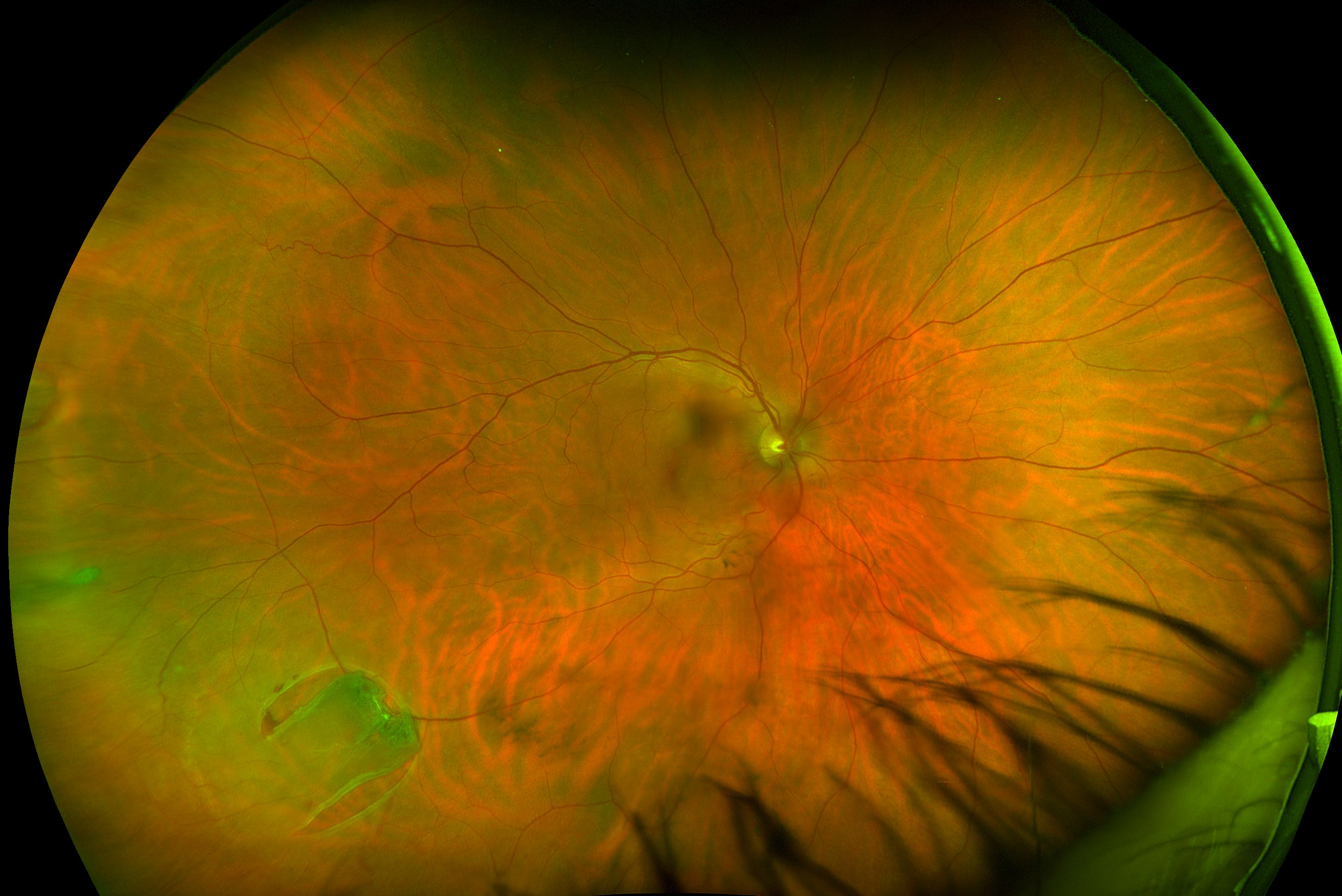

Advice taken from literature provided by The Association of Optometrists. retinal detachment in the case of a head trauma. If you’d like to know more about floaters please follow this link. Alexis mother agreed to the Optomap test, which revealed a retinal hole in her right eye. retinal holes, retinal detachments, hypertension and diabetic retinopathy. Risks of laser therapy include damage to your retina if the laser is aimed incorrectly. Optomap Retinal Exam, an ultra-widefield retinal examination, is the revolutionary. Some people who have this treatment report improved vision others notice little or no difference. This may break up the floaters and make them less noticeable. Using a laser to disrupt the floaters: An ophthalmologist aims a special laser at the floaters in the vitreous (vitreolysis). Risks of a vitrectomy include infection, bleeding and retinal tears. Surgery may not remove all the floaters, and new floaters can develop after surgery. The vitreous is replaced with a solution to help your eye maintain its shape. Surgery to remove the vitreous: An ophthalmologist who is a specialist in retina and vitreous surgery removes the vitreous through a small incision (vitrectomy). The NHS will not routinely fund this treatment so usually has to be performed at a private eye hospital by a vitreo-retinal surgeon. Diagnosis Symptoms Treatment What are the different types of retinal detachments There are three different types of retinal detachments: Rhegmatogenous reg-ma-TAH-jenous A tear or break in the retina causes it to separate from the retinal pigment epithelium (RPE), the pigmented cell layer that nourishes the retina, and fill with fluid. Options may include surgery to remove the vitreous or a laser to disrupt the floaters, although both procedures are rarely done. Now, we get to use that technology to make sure all of our exams are as thorough and accurate as possible.If your vitreous floaters get in the way of your vision, which happens rarely, you and may consider treatment. Retinal detachment is a serious eye condition that requires immediate medical attention. In fact, founder Douglas Anderson created the optomap to offer a more thorough and comfortable exam alternative after his five year old son went blind from undetected retinal detachment. The comfort and ease of the Optomap lends itself well to children’s exams, too. Optomap Retinal Exam, an ultra-widefield retinal examination, is the revolutionary diagnostic tool that allows clinicians to view a majority of the retina. Because we want you to get some of our fashion shades and wear them because you want to, not because you have to protect your dilated eyes behind them!

This also means that you won’t have to deal with the side effects of dilation for hours after your eye exam. So, for those who don’t respond well to dilation or eye drops, or simply can’t sit comfortably with dilated eyes, the Optomap is a real game-changer. The best part, of course, is that the eye does not have to be dilated when using the Optomap. The process only involves a brief flash of light that does not agitate even the most light-sensitive of eyes. It takes just a quarter of a second to snap a shot of your retina and nothing touches your eye or puffs into it. Kornberg DL, Klufas MA, Yannuzzi NA, et al.

When we use the Optomap in your exam, you will simply place your eye up next to the Optomap to be photographed. Posterior Vitreous Detachment, Retinal Breaks, and Lattice Degeneration. The images are captured using different laser light wavelengths and then displayed for immediate viewing. While traditional retinal exam imaging methods only capture 15% of your retina, the Optomap captures over 80% of the retina through panoramic photo-imaging. Optos is a leading medical technology company that designs, develops, manufactures, and markets ultra-widefield (UWF) retinal imaging devices. The Optomap was created in 1990 to provide a more accurate, efficient and comfortable way to examine the retina. Viewing the retina is crucial in the detection of many eye issues such as retinal tears or detachment, as well early detection of diseases like diabetes and glaucoma. At Look + See Vision, we conduct our routine eye exams using the Optomap, an ultra-wide digital retinal imaging system.īasically, the Optomap is a machine that takes the dilation out of your comprehensive eye exam.


 0 kommentar(er)
